Dr Partl
Dr Partl
Address
Hornweg 28, 6370 Kitzbühel
GPS
47.44965597938, 12.39237028911
Telephone
Dr Partl
People are our focus – working with my fantastic team is a joy. With in-depth training, experience, and state-of-the-art equipment, we create transparency and clarity to quickly alleviate any fears and answer our patients’ questions.
Dr. Eberhard Partl
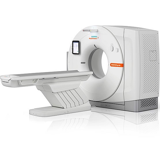
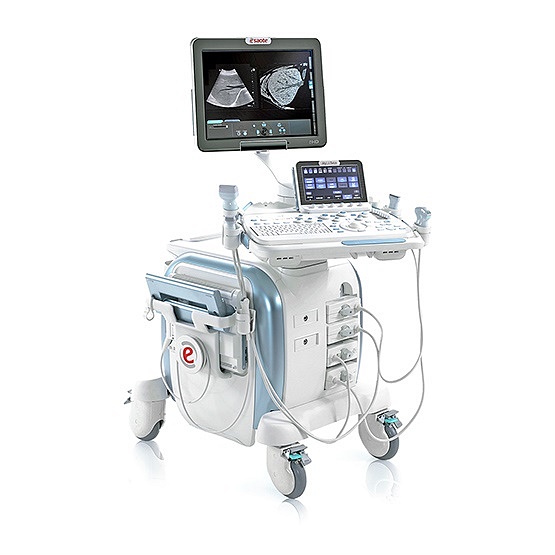
MRI 3t
 Paid service of the institute
Magnetic resonance imaging
of all joints, spine
, intervertebral
discs ,
neurological examinations of the skull
, angiography (vascular examinations),
organ screenings (e.g. prostate)
, note:
No reimbursement is possible through statutory health insurance!
Siemens 3T MAGNETOM Skyra
The 3T MAGNETOM Skyra is Siemens’ most powerful MRI scanner and the only one of its kind in Tyrol outside of Innsbruck University Hospital. Its high magnetic field strength of 3 Tesla (most scanners operate at 0.35–1.5 Tesla) and the latest technologies enable the best image quality in the shortest examination times, as well as special diagnostic techniques that deliver precise images of even the smallest vascular changes. Further advantages: The generous internal diameter of 70 cm and a low noise level make the examination even more comfortable.
Precise image sharpness for the highest diagnostic quality. Magnetic resonance imaging (MRI) allows the structures and functions of internal organs to be displayed in high detail and pathological changes to be precisely identified. It is particularly suitable for examining soft tissue such as muscles and ligaments. Unlike CT and X-ray examinations, no X-rays are used. Because we have two state-of-the-art scanners, you can get an appointment quickly.
computed tomography
 Paid service of the institute
CT (computer tomography)
Skull
Paranasal sinuses
Neck organs
Internal organs
Thorax (chest cavity)
Abdomen (abdominal cavity)
Spine
CT special examinations
Dental CT
Low-dose lung CT (lung screening)
Calcium scoring
Angiography (3D vascular imaging)
Note
: We are accepted as a private practice by the statutory Tyrolean health insurance companies – i.e. a portion of the costs (up to 80%) are refunded.
Computed tomography (CT) provides complex and precise insights. In this examination, a unit consisting of an X-ray tube and a detector opposite it rotates in fractions of a second around the patient, who is slowly moved through the center of the scanner while lying down. This produces cross-sectional images of the region under examination, from which reconstructions can then be performed at different levels. This creates precise images of internal organs, bones, and blood vessels.
digital x-ray
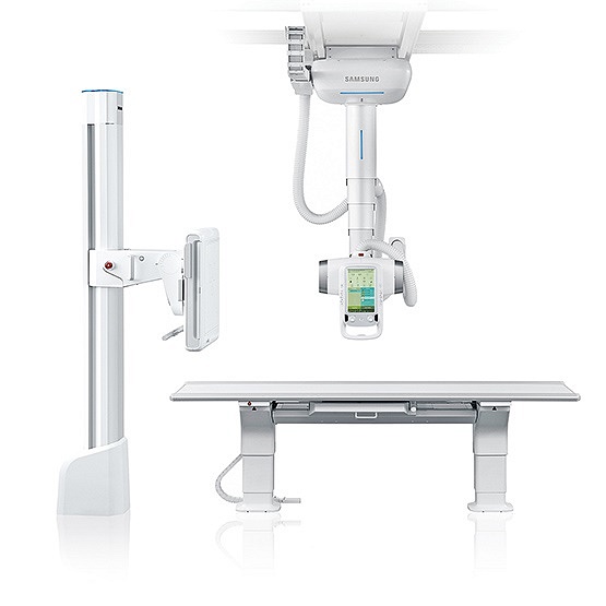 Free service from the practice
Digital X-rays allow targeted diagnoses for:
Suspected fractures/dislocations
Joint problems (osteoarthritis, rheumatism)
Spine and leg full-body radiographs
Intestinal obstruction, tumor
screening (e.g., lung X-ray)
Bone age determination in children
Less harmful than traditional X-rays – because fewer rays are released. X-rays are electromagnetic waves that penetrate the body’s organs and create images. Such examinations are performed while the patient is sitting, lying down, or standing and take only a few minutes. We work exclusively with digital X-ray machines, as these X-ray images are available immediately in the highest quality and can be post-processed on a computer – for example, to better identify specific structures.
digital fluoroscopy
ultrasonic
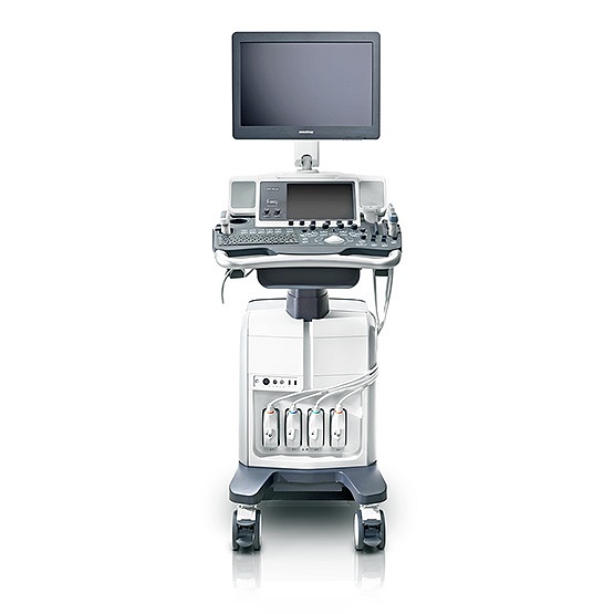 Free service from the practice
Ultrasound (sonography) for pain or as part of preventive care:
Soft tissues of
the neck Thyroid
Lymph nodes
Breast
Abdominal organs
All soft tissue regions
For examination of:
Arteries
Veins
Organ blood flow
Simple, convenient, and all on screen in less than 15 minutes: An ultrasound examination can be used for virtually all organs. Whether for preventative screening or to diagnose acute conditions, a sonography is radiation-free and comfortable – usually while lying down. During this imaging procedure, a gel-covered ultrasound probe is moved over the body region to be examined. The sound waves are reflected in different ways, creating a real-time image of the body’s interior. This allows us to make precise diagnoses – completely stress-free for you.
mammography with tomosynthesis
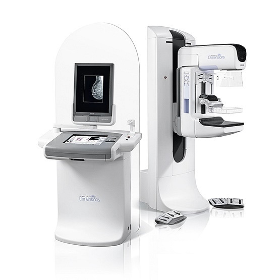 Free service from the practice
Mammography
Breast cancer screening from the
age of 40
In case of pain or palpable findings
Annual check-ups if there is a positive family history (breast cancer in the family)
Breast sonography (ultrasound examination of the breast)
In case of pain or palpable findings
In case of dense breasts (mastopathy)
To exclude inflammation or tumors
Tomosynthesis (slice-by-slice imaging of the breast) for the early detection of breast tumors. Breast cancer (mammary carcinoma) is a malignant change in breast tissue – the most common cancer in women in Austria. Regular screening examinations for early detection are therefore important. From the age of 40, we recommend mammography screening, which we perform in our practice. This is a particularly accurate method in which the breast is examined using special X-ray technology and ultrasound. Please bring any existing mammograms with you for the examination – they make it easier to monitor changes in the breast. Dr. Partl’s X-ray practice has been awarded the Mamma Certificate by the Federal Radiology Association and the Austrian Radiological Society. All mammograms are double-read by a suitably trained and certified radiologist.
vascular Doppler
 Free service from the practice
When it comes to your blood vessels, everything must be clear: Color-coded Doppler or duplex sonography (also called vascular Doppler) is one of the routine examinations used to diagnose vascular diseases. This special ultrasound examination measures the blood flow velocity in arteries and veins and assesses their diameter and condition. This allows for the detection of narrowing and circulatory disorders. For example, it can be used for stroke prevention, to detect thrombosis early, or to monitor blood flow to organs after surgical procedures.
bone density measurement
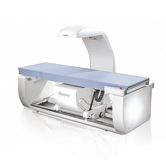 Paid service of the institute
dexa bone density measurement in the case of:
Clinical suspicion of osteoporosis
Osteoporosis screenings during menopause
Follow-up checks for osteoporosis/therapy control
Suspicion of insufficient mineral absorption
Complaints such as bone pain, hunchback, etc.
Ongoing cortisone therapy
Note
: We are accepted as a private practice by the statutory Tyrolean health insurance companies – i.e. a portion of the costs (up to 80%) are refunded.
Transparent osteoporosis diagnosis – with DEXA bone density measurement. This involves determining the bone mineral content in the lumbar spine and femoral neck using a low dose of radiation: The lower the mineral content, the higher the risk of bone fractures. The examination takes approximately 10 minutes, during which you lie quietly on a special examination table. First, the lumbar spine (L1 to L4) and hip are measured. We then evaluate the obtained data using a special computer program and present it to you in a diagram.
OPENING HOURS
Monday
07:30- 16:00
Tuesday
07:30- 14:00
Wednesday
07:30- 14:00
Thursday
07:30- 12:00 + 16:00 – 19:30
Friday
07:30- 12:00
Saturday
closed
Sunday
closed